Describe the Equipment Used in Extraoral Imaging
DENTAL RADIOGRAPHYpdf from SCHOOL OF BMI14B at KPJ University College Nilai. Tell me when Its Available.
Extraoral Radiography
Extra oral Radiography.
. Equipping operatories with portable extraoral aerosol extractors can improve the safety of care by helping contain dental bioaerosols. Describe the equipment used in extraoral imaging. This is an alternate ISBN.
To aid in patient positioning and alignment of the x-ray beam special head positioning and beam alignment devices can be added to intraoral x-ray machines. It constitutes of Two-dimensional digital array providing an area detector fixed on a rotating gantry A three- dimensional 3D cone shaped x-ray beam. In some cases an extraoral film is used because the patient has swelling or severe pain and is unable to tolerate the placement of intraoral films.
The extraoral radiograph provides an overall image of the skull and jaws. 2Special head positioning and beam alignment devices can be added. Customize your workflow with open architecture which allows for seamless integration with most third-party products.
Fewer intraoral pieces of equipment are used when taking extraoral radiographs such as panoramic and cephalometric films. Apicoectomy and obturation of the apex a bold act. Extraoral radiographs are used for the following reasons.
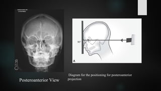
A special device used to hold the intensifyin. View the primary ISBN for. The lateral cephalometric projection is used to evaluate facial growth and development trauma disease and developmental abnormalities.
The use of covers over the bite guide is less desirable23 Table 6. Periapical Radiograph Locurcio LL Leeson R. A standard intraoral x-ray machine may be used for some extraoral images.
Modern Dental Assisting 10th Edition. To prepare the patient for panoramic imaging explain the procedure to the patient as him or her to remove all metal objects from head and. G screen and the extraoral film is available in various sizes that correspond the.
The authors have no commercial conflicts of interest to disclose. View the primary ISBN for. Null null Edition Textbook Solutions.
Extraoral images are particularly beneficial for patients requiring orthodontic treatment dental implants and oral surgical procedures. Medium or high speed rare-earth screenfilm combination are recommended to reduce pts exposure. Describe the equipment used for extraoral imaging.
Most extraoral exposures use screen film place in cassette that has an intensifying screen grid a device used to reduce the amount of scatter radiation that reaches extraoral film during exposure. Created using patented software-driven SCARA technology Planmeca ProMax 3D provides dependable 2D3D imaging with versatile programs and volume sizes to accommodate a wide range of clinical needs. List the steps for patient preparation and positioning in panoramic imaging.
Our equipment is designed to promote proper posture for doctors assistants and patients alike while helping to provide the workflow that best suits your practice needs. Doni L Bird Debbie S Robinson Rent Buy. Midmark Extraoral Imaging System.
An imaginary horseshoe shaped area used for jaw placement what are advantages of extraoral radiographs minimize radiation to the patient provide an overview of the skull and jaws and prevent the patient from gagging. This is an alternate ISBN. The equipment used in panoramic imaging are the panoramic x-ray unit screen film intensifying screens and cassette.
Cephalometric and skull views require at least a 20 25 cm 8 10 inch image receptor whereas oblique lateral projections of the mandible can be obtained with a 13 18 cm 5 7 inch image receptor. The extraoral films are used in combination with the cassettes and the intensifying screens. Midmark Extraoral Imaging System 2D Panoramic with Cephalometric X-Ray Midmark Extraoral Imaging System 2D Panoramic X-Ray Midmark Extraoral Imaging System 3D X.
1A standard intraoral x-ray machine may be used for a variety of extraoral projections. The equipment used in extra oral imaging are the x-ray unit film intensifying screens cassette and grid. Typical extraoral x-ray images include panoramic cephalometric and cone beam computed tomography CBCT projections.
A lateral cephalograph is a sagittal projection of the skull that includes both the hard and soft tissues. First because so much data is captured in a single X-ray done right onsite diagnoses are quicker and more accurate. This 2 credit hour self-study activity is electronically mediated.
A case of periradicular surgery. Metallic letters L R are used to determine left right sides of pt as well as grids to reduce the fog. Bite guides should be sterilized or be single-use disposable types.
The chances of contamination with patient blood or saliva are greatly reduced. Having a cone beam also attracts new patients who expect a highly skilled doctor using the latest techniques. They provide you with more detail of structures around particular teeth.
PRINCIPLE- Cone-beam scanners use a two- dimensional digital array providing an area detector rather than a linear detector as CT does. IMAGING TECHNIQUE II DENTAL RADIOGRAPHY LECTURER Medical Imaging Department KPJ Healthcare University. This leads to more efficient patient treatment in fewer visits.
Extraoral radiographs are produced with conventional dental x-ray machines certain models of panoramic machines or highercapacity medical x-ray units. A 3D unit also allows general dentistry practices to. Extraoral film equipment.
All extra oral radiographic projections should be performed using screen film. Null null Edition Textbook Solutions. Planmeca ProMax 3D Imaging Units.
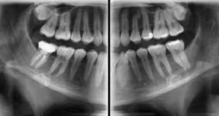
To identify trauma or fractures. Periapical around the apex radiographs are usually used to assess specific teeth. This course was published in the November 2021 issue and expires November 2024.
Identify the specific purpose of each of the extraoral film projections. Provide the highest level of care and get the most efficiencies from your workflows with 2D and 3D technology. 3The panoramic tubehead is used in conjunction with a special extension arm and a device known as a cephalostat or craniostat.
Ad Browse Our Large Selection of 3D Imaging Machines From Industry Leading Manufacturers.

Examples Of Intraoral And Extraoral Bitewing Radiographs A Intraoral Download Scientific Diagram
Extraoral Radiography
Extraoral Radiograph Lecture
Everything You Need To Know About Extraoral Bitewings Renew Digital Llc
No comments for "Describe the Equipment Used in Extraoral Imaging"
Post a Comment